
Stanford-SLAC Cryo-EM Center Facilities & Resources

S2C2 Cryo-EM Facilities
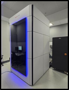
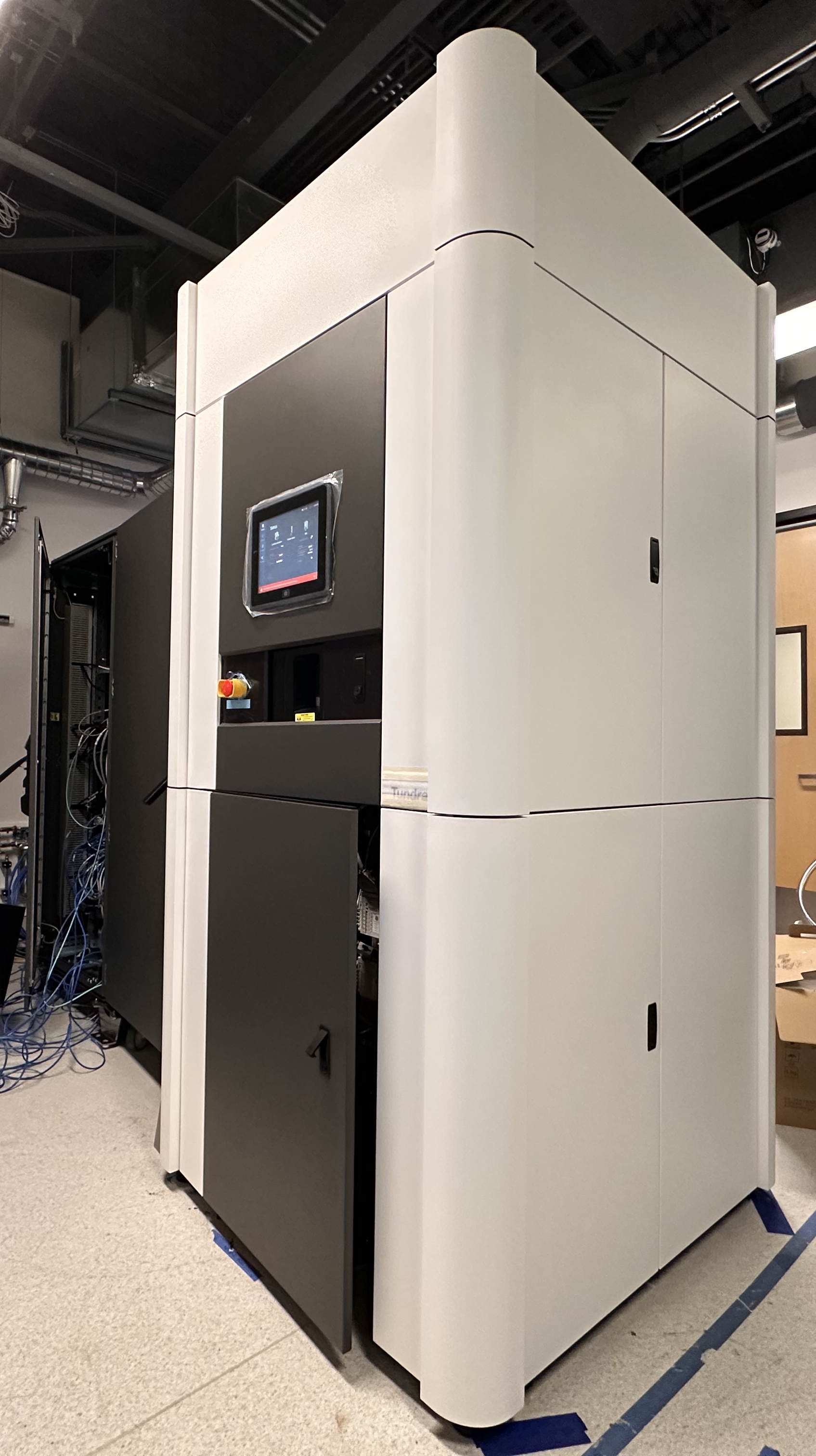
S2C2 is equipped with cutting-edge instrumentation. Our facility boasts four state-of-the-art 300 keV electron microscopes (Titan Krios) manufactured by Thermo Fisher Scientific (named as Alpha, Beta, Gamma and Delta), each with various gun sources, electron energy filters, and detectors, ensuring versatility for your research needs. Additionally, we have a 200 keV electron microscope (Glacios 2) and a 100 keV electron microscope (Tundra). All dedicated to high-resolution data collection, training, and specimen screening. Furthermore, our facility is accessible to other Titan Krios electron microscopes (TEM2) if needed. To support your experiments, we provide a range of sample vitrification devices, including Vitrobot, Leica EM GP2 plunger, Chameleon, Vitrojet, and Gatan CP3 plunger. To minimize ice contamination, some of these devices are located in a low humidity room.
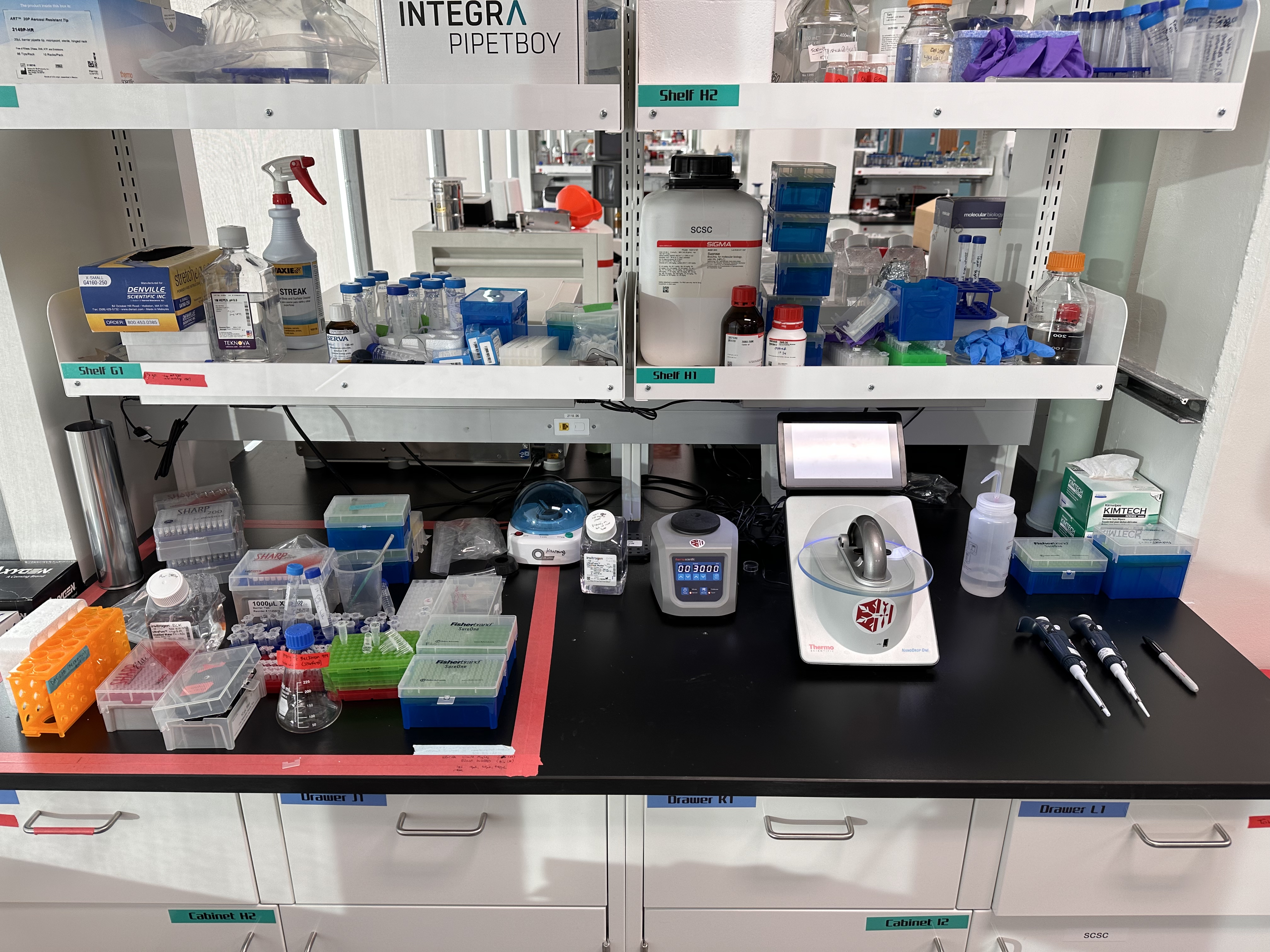
For the final step in preparing fresh samples before vitrification, users have access to biochemical preparation equipment. It's important to note that our CryoEM facilities are authorized to handle BSL2 samples with proper approval from the Stanford Biosafety office.

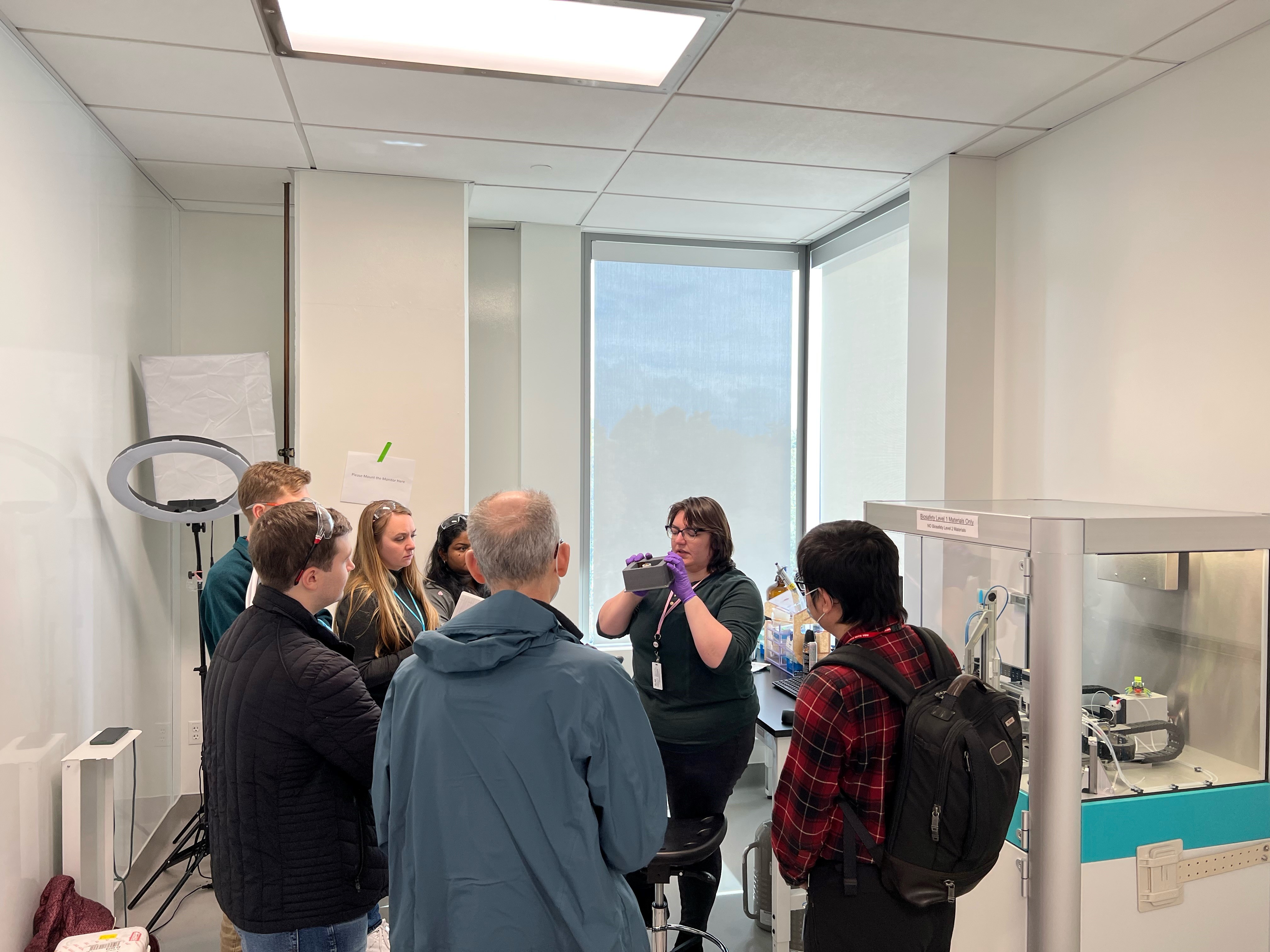
S2C2 Workshop for Cryogenic Sample Preparation

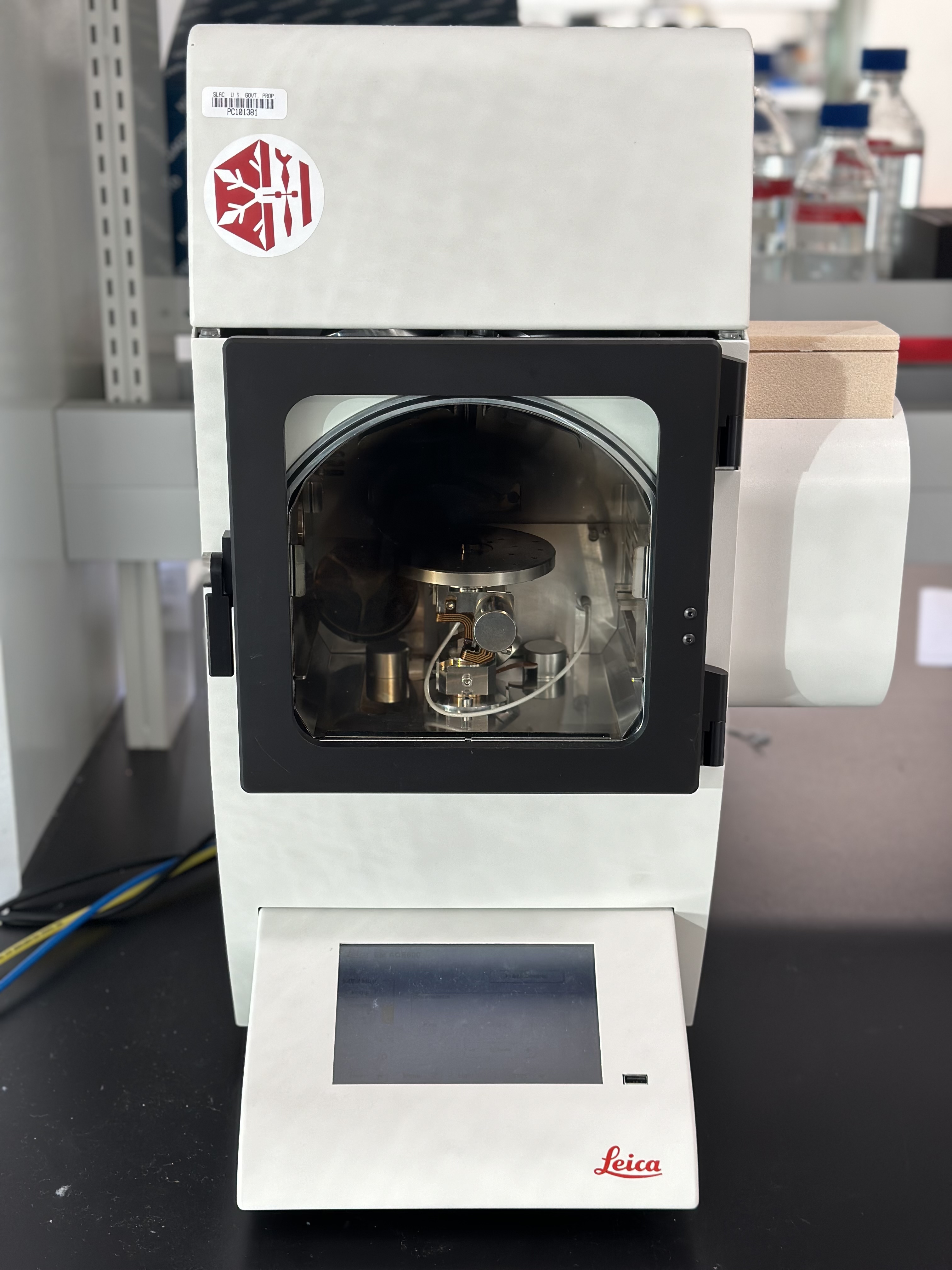
S2C2 Workshop for Cryogenic Sample Preparation: Take advantage from the automatic, blotless grid technology and high-speed plunging of Chameleon to optimize your sample prep.

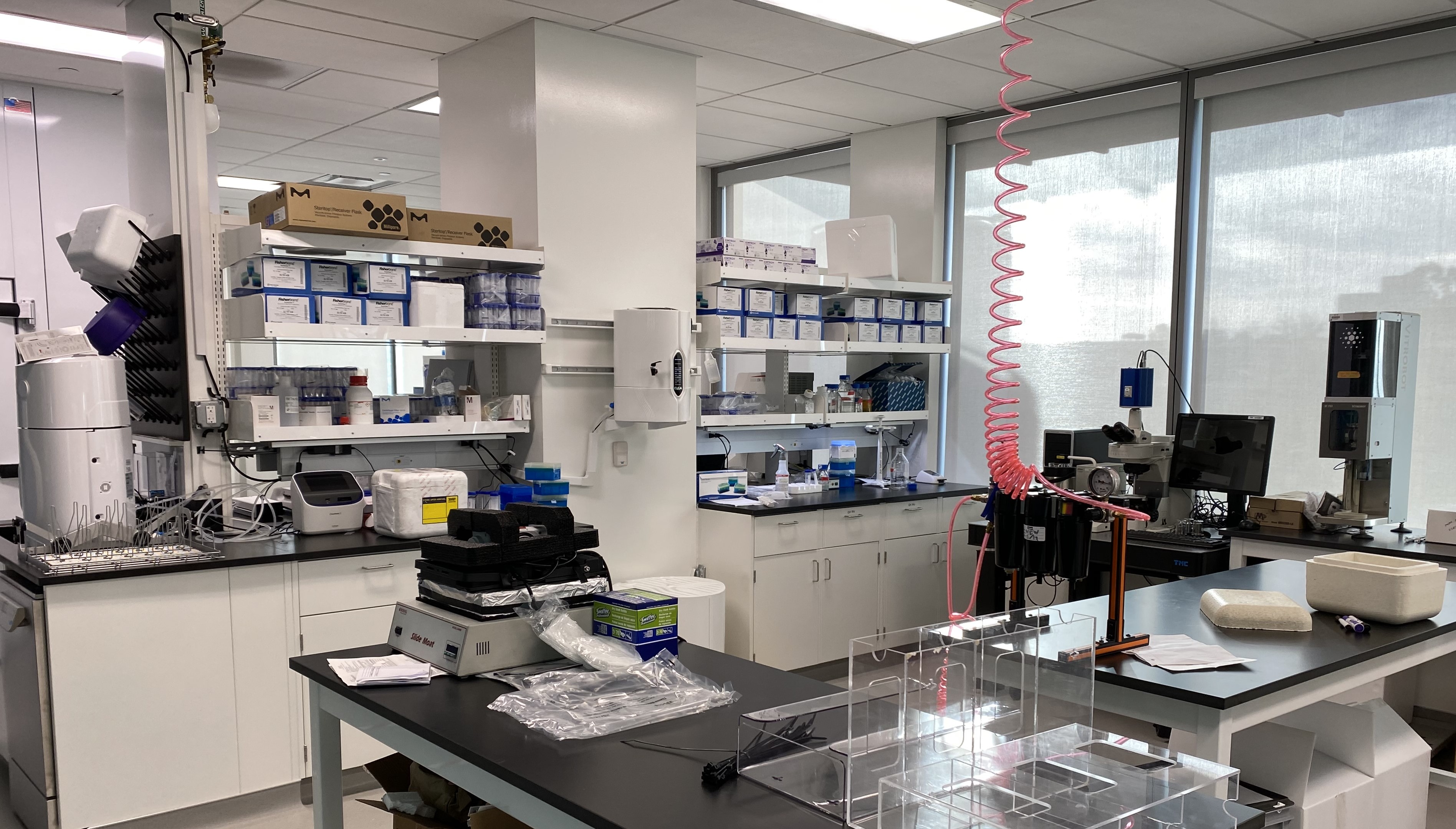
CryoEM Wet Lab Bldg. 057 Arrillaga Science Center (ASC)

CryoEM Wet Lab - Bldg. 57 Arrillaga Science Center: Equipped with a complete infrastructure of equipment available for the preparation of your samples before freezing.


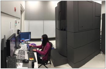
Titan Krios G3i (TEM-Alpha)

Titan Krios G3i (TEM-Alpha): Stanford Grad Student, Rachael Kretsch, working on the 300keV Cold FEG and Fringe-Free Imaging microscope with Selectris X energy filter and Falcon 4i detector, ideal for CryoEM SPA and Tomography analyses.

Titan Krios G3i (TEM-Beta)

Titan Krios G3i (TEM-Beta): Stanford Grad Student, Lily Xu working on the 300keV X-FEG and Fringe-Free Imaging microscope with BioQuantum energy filter and K3 detector, ideal for CryoEM SPA (EPU Multigrid software) and Tomography analyses.

Titan Krios G3i (TEM-Gamma)

Titan Krios G3i (TEM-Gamma): 300keV Cold FEG and Fringe-Free Imaging microscope with Selectris X energy filter, Falcon 4i and Ceta-D detectors, ideal for CryoEM SPA, Tomography and Micro-ED analyses.

Titan Krios G4 (TEM-Delta)

Titan Krios G4 (TEM-Delta): 300keV Cold FEG and Fringe-Free Imaging microscope with Selectris X energy filter and Falcon 4i detector, ideal for CryoEM SPA and Tomography analyses.

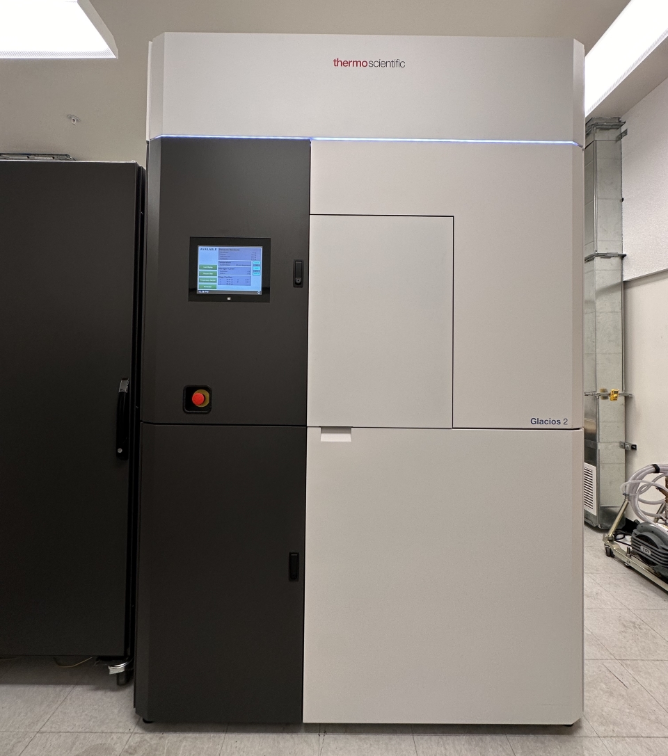
Talos Glacios 2

Talos Glacios 2: 200keV X-FEG and Fringe-Free Imaging microscope with Selectris X energy filter, Falcon 4i and Ceta-D detectors, ideal for CryoEM SPA, Tomography and Micro-ED analyses.
photo taken by Dr. Alexandre Cassago

Talos Tundra

Talos Tundra: 100keV X-FEG microscope with Falcon C and DE Centurion detectors, ideal for CryoEM SPA.
photo taken by Dr. Alexandre Cassago

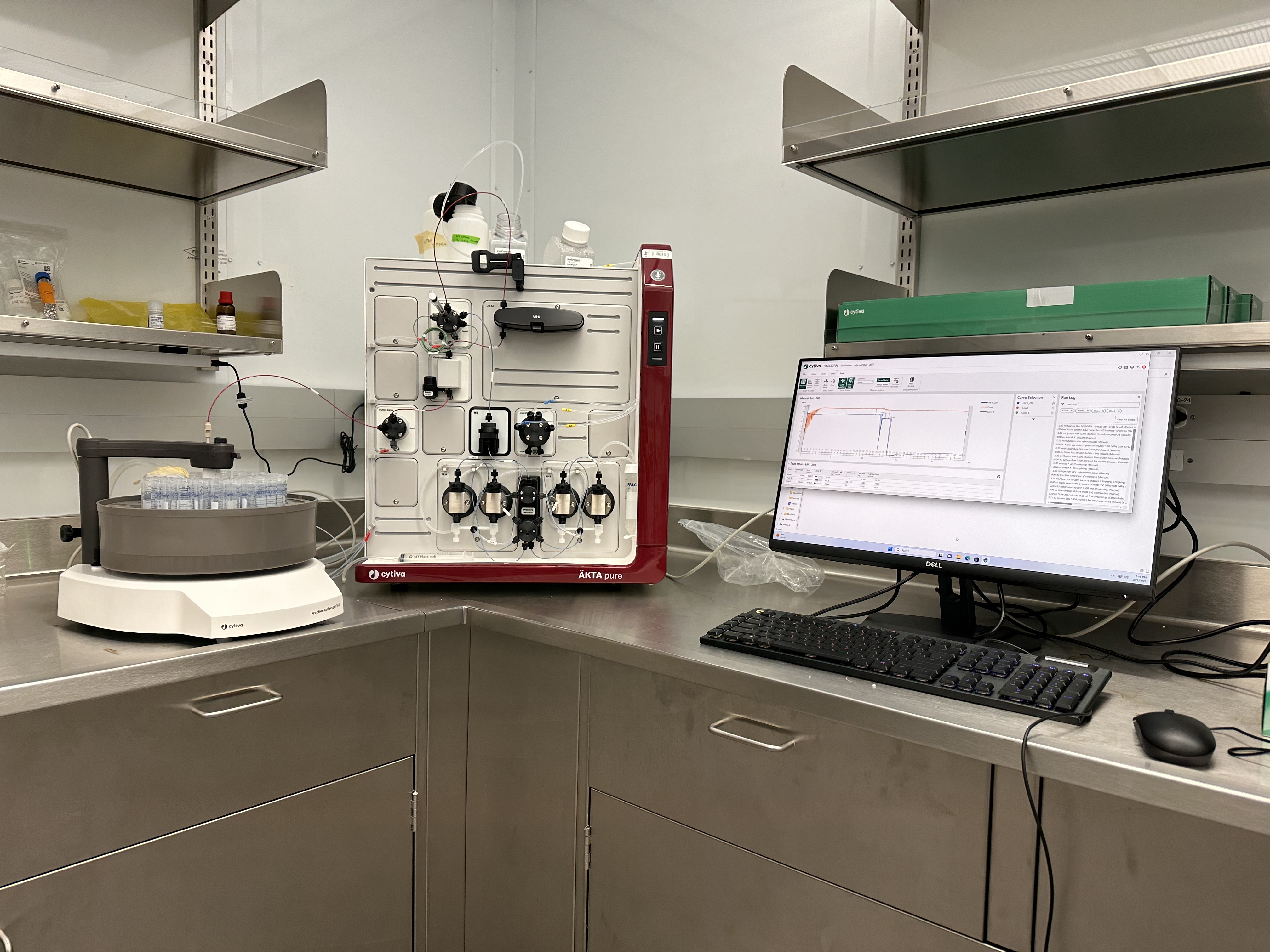
ÄKTA Chromatography Systems

ÄKTA Chromatography Systems: The most modern and user-friendly equipment for protein purification and sample preparation before freezing.
photo taken by Dr. Alexandre Cassago

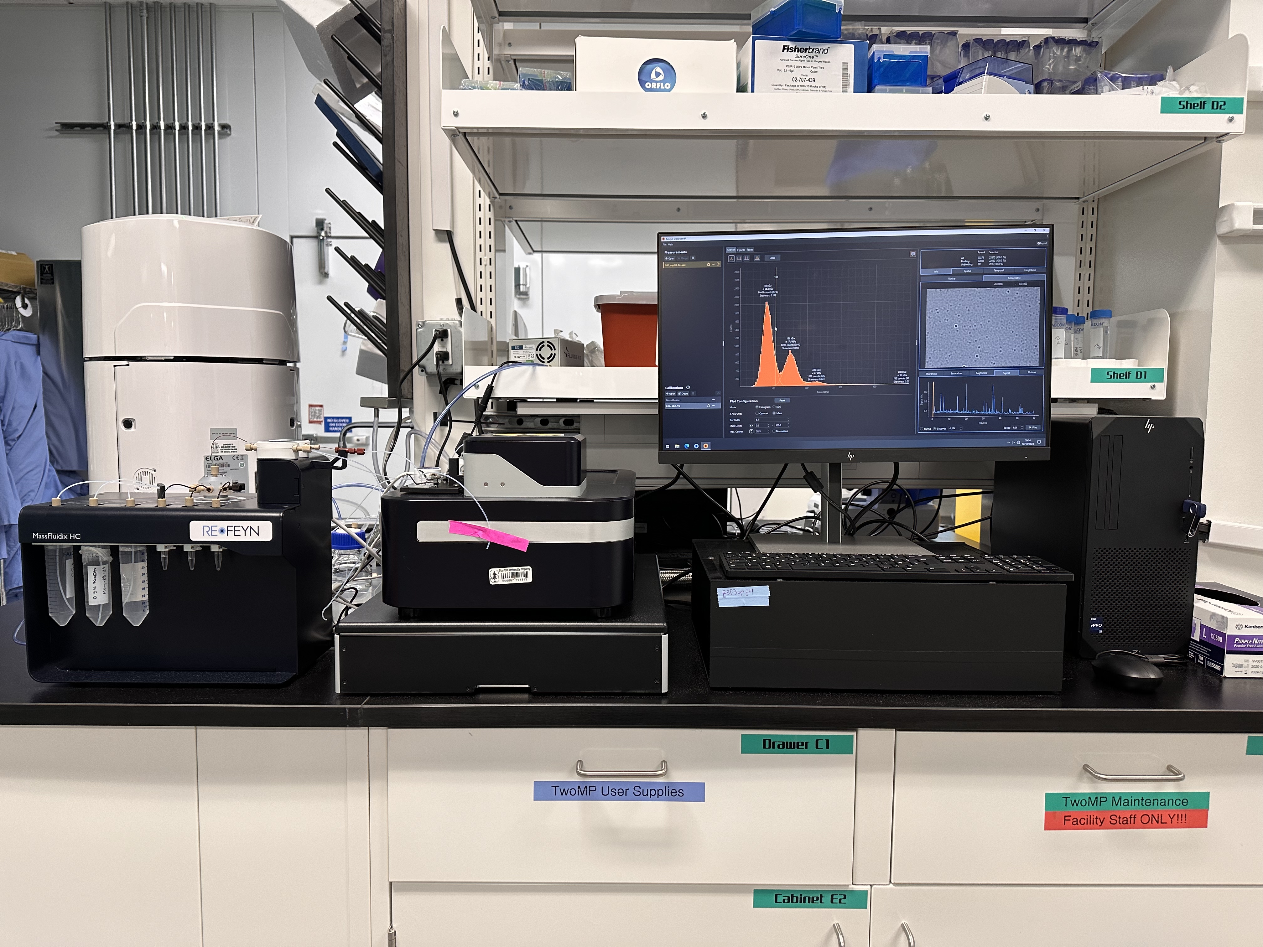
Refyn Mass Photometer

Refyn Mass Photometer: Mass photometry with a particle mass range between 30 kDa and 5 MDa from proteins, antibodies, and nucleic acids. Ideal for determining the oligomeric state and protein-ligand interactions of your sample.
photo taken by Dr. Alexandre Cassgo

CryoEM Wet Lab - Bldg. 57 Arrillaga Science Center

CryoEM Wet Lab - Bldg. 57 Arrillaga Science Center: Equipped with RNA Workstation Area for preparing RNA samples.
photo taken by Dr. Alexandre Cassago

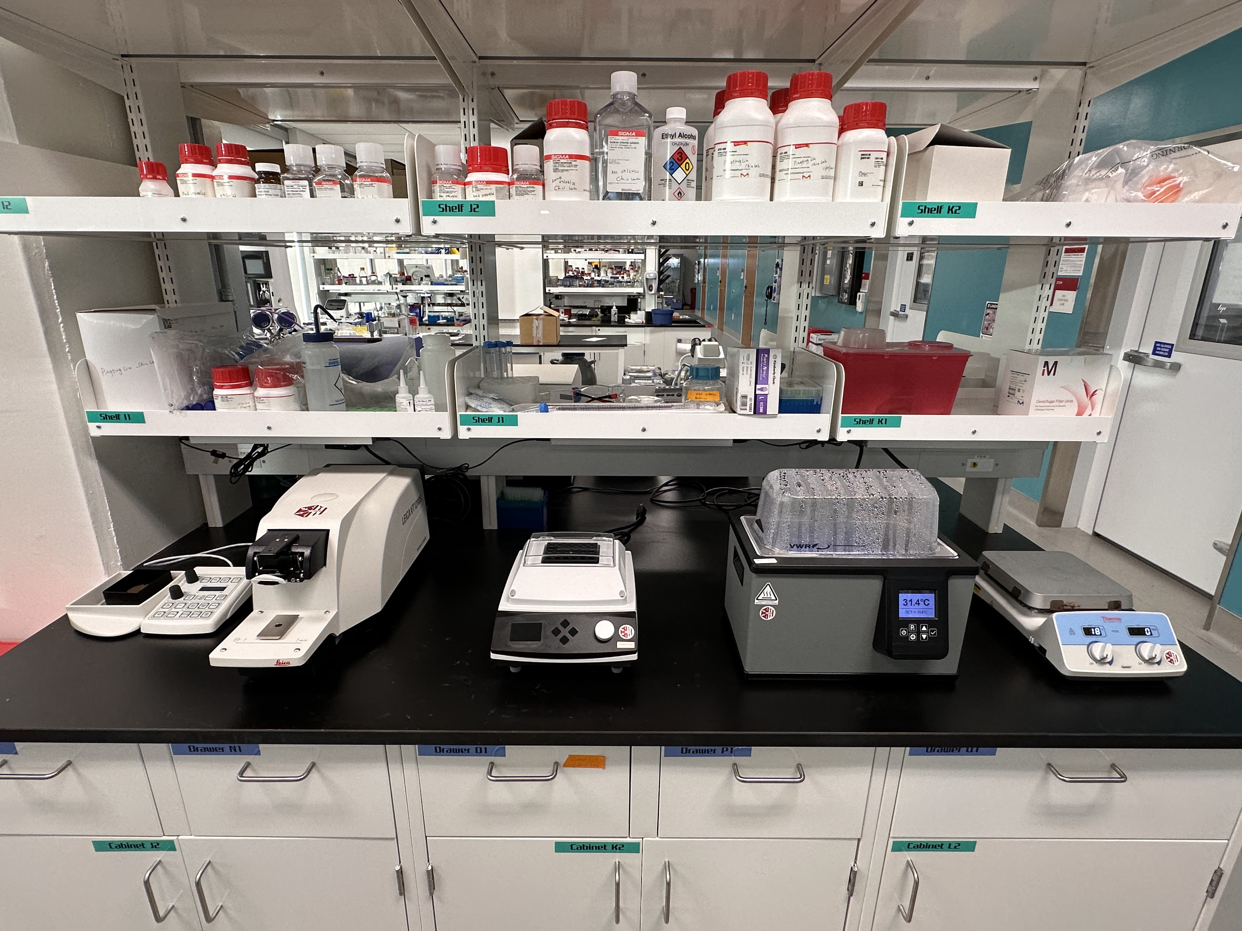
CryoEM Wet Lab - Bldg. 57 Arrillaga Science Center

CryoEM Wet Lab - Bldg. 57 Arrillaga Science Center: Equipped with semi-automated Leica VT1200 Vibratome and Thermal Bath.
photo taken by Dr. Alexandre Cassago

CryoEM Wet Lab - Bldg. 57 Arrillaga Science Center

CryoEM Wet Lab - Bldg. 57 Arrillaga Science Center: Equipped with EM ACE600 Sputter Coater for fine grained thin films preparation for electron microscopy prep.
photo taken by Dr. Alexandre Cassago
| Microscope | Alpha | Beta | Gamma | Delta | Glacios 2 | Tundra | TEM2 |
|---|---|---|---|---|---|---|---|
| Model | Titan Krios G3i | Titan Krios G3i | Titan Krios G3i | Titan Krios G4 | Glacios G2 | Tundra | Titan Krios G3 |
| Imaging Software | EPU | EPU | EPU | EPU | EPU | Smart-EPU | EPU |
| Cold FEG | Yes | No | Yes | Yes | No | No | No |
| Fringe Free | Yes | Yes | Yes | Yes | Yes | No | Yes |
| Energy Filter | Selectris X | BioQuantum | Selectris X | Selectris X | Selectris | N/A | BioContinuum |
| Detector(s) | Falcon 4i Ceta | K3 | Falcon4i | Falcon 4i | Falcon 4i | Falcon C | K3 |
| Apoferritin Performance | 1.27 Å | 1.34 Å | 1.46 Å | 1.33 Å | 1.9 Å | 2.50 Å | 1.7 Å |
Data Management and Computational Resources
All experimental data collected under S2C2 will be held on disk for two months from the day of collection. The data is transferred as soon as it is collected and is therefore immediately available for download via either the usual unix tools (rsync, scp, bbcp, etc) or through globus. More details regarding data transfer can be found at https://confluence.slac.stanford.edu/x/mYoYDg.
After two months, the data is then moved onto tape and will be kept for 22 additional months. Requests for data retrieval after the data is moved onto tape can be made via email to unix-admin@slac.stanford.edu with specific information regarding the date of the data collection, the proposal number and the microscope that was used.
Two years after the data is collected, the data will be purged completely from our systems.
As part of our data services, all collected movie images will be automatically aligned (motion corrected) and CTF calculations performed and reported.
